Monthly Archives: Nisan 2018

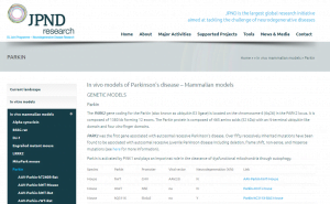
JPND’s database of experimental models for Parkinson’s disease has recently been updated with 29 additional new pages on in-vivo mammalian models.

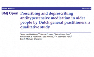
The database,  which was first made available to the community in 2017, is a unique collection aimed at presenting and fostering scientific discussion around the experimental models currently available to study Parkinson’s disease.

The database is continuously updated to reflect developments from the scientific literature and is a major effort by JPND to help researchers in the field. Together with an exhaustive description of each model and the studies that have already been done, there is a section for comments. You are welcome to add your comments to any of the models you have had experience with or to present alternative models.

We hope that you will join this growing community of scientists. To consult the database and add your contributions, please visit: https://www.neurodegenerationresearch.eu/models-for-parkinsons-disease/.

If you’d like any additional information, don’t hesitate to contact us directly at experimentalmodels@jpnd.eu.


ALS and frontotemporal dementia (FTD) are two neurodegenerative diseases with a toxic relationship, according to a new paper published in Nature Medicine. The study describes how a mutation in a gene, called C9ORF72, leads to toxicity in nerve cells—causing 10 percent of all cases of ALS, and an additional 10 percent of FTD.

To understand how this happens, the researchers extracted blood from ALS patients carrying the C9ORF72 mutation, and reprogrammed these blood cells into the motor nerve cells that degenerate and die in the disease. They also extracted blood from healthy patients, reprogrammed these blood cells into motor nerve cells, and used gene editing to delete the C9ORF72 gene.

Whether patient-derived or gene-edited, all motor nerve cells with the mutation had reduced amounts of the protein normally made by the C9ORF72 gene. Furthermore, by adding supplemental C9ORF72 protein, the researchers could stop the motor nerve cells from degenerating.

Through a series of experiments, the researchers revealed that the motor nerve cells use C9ORF72 protein to build lysosomes—which are cellular compartments used to engulf and break down toxic proteins and other garbage.

Without enough lysosomes, the cells accumulate two key types of garbage. The first type is a large, toxic protein produced by the mutated C9ORF72 gene itself. The second type is an excessive number of receptors, or molecules that receive signals from a neurotransmitter known as glutamate. These receptors respond to glutamate by causing the motor nerve cell to activate. Too much activation can kill a motor nerve cell.

The researchers are now using patient-derived motor nerve cells to test potential drugs—with a focus on those that affect lysosomes.

Paper: “Haploinsufficiency leads to neurodegeneration in C9ORF72 ALS/FTD human induced motor neurons”

Reprinted from materials provided by the Keck School of Medicine at USC.

A team has developed a system to model Huntington’s in human embryonic stem cells for the first time. In a report published in Development, they describe early abnormalities in the way Huntington’s neurons look, and how these cells form larger structures that had not previously been associated with the disease.

Huntington’s is one of the few diseases with a straightforward genetic culprit: One hundred percent of people with a mutated form of the Huntingtin (HTT) gene develop the disease. The mutation takes the form of extra DNA, and causes the gene to produce a longer-than-normal protein.

Research on Huntington’s has thus far relied heavily on animal models of the disease. Suspecting that the disease works differently in humans, the researchers developed a cell-based human system for their research. They used the gene editing technology CRISPR to engineer a series of human embryonic stem cell lines, which were identical apart from the number of DNA repeats that occurred at the ends of their HTT genes.

When cells divide, they typically each retain one nuclei. However, some of these mutated cells flaunted up to 12 nuclei—suggesting that neurogenesis, or the generation of new neurons, was affected.

Treatments for Huntington’s have typically focused on blocking the activity of the mutant HTT protein. However, this research shows that the brain disruption may actually be due to a lack of HTT protein activity. The researchers created cell lines that completely lacked the HTT protein. These cells turned out to be very similar to those with Huntington’s pathology, corroborating the idea that a lack of the protein—not an excess of it—is driving the disease.

Article:  “Chromosomal instability during neurogenesis in Huntington’s disease.”
Reprinted from materials provided by Rockefeller University.

People with Alzheimer’s disease are known to have disturbances in their internal body clocks that affect sleep/wake cycle and may increase their risk of developing the disorder. Now, new research published in JAMA Neurology indicates that such circadian rhythm disruptions also occur much earlier in people whose memories are intact but whose brain scans show early, preclinical evidence of Alzheimer’s disease.

Previous studies conducted in people and in animals have found that levels of amyloid fluctuate in predictable ways during the day and night. Amyloid levels decrease during sleep, and several studies have shown that levels increase when sleep is disrupted or when people don’t get enough deep sleep.

The researchers tracked circadian rhythms in 189 cognitively normal, older adults with an average age of 66. Of the participants, 139 had no evidence of the amyloid protein that signifies preclinical Alzheimer’s. Most had normal sleep/wake cycles, although several had circadian disruptions that were linked to advanced age, sleep apnea or other causes.

But among the other 50 subjects — who either had abnormal brain scans or abnormal cerebrospinal fluid — all experienced significant disruptions in their internal body clocks, determined by how much rest they got at night and how active they were during the day. Disruptions in the sleep/wake cycle remained even after the researchers statistically controlled for sleep apnea, age and other factors.

By tracking activity during the day and night, the researchers could tell how scattered rest and activity were throughout 24-hour periods. Subjects who experienced short spurts of activity and rest during the day and night were more likely to have evidence of amyloid buildup in their brains, the researchers said.

Paper: “Circadian Rest-Activity Pattern Changes in Aging and Preclinical  Alzheimer Disease”
Reprinted from materials provided by Washington University School of Medicine.

A new study published in Scientific Reports shows that low levels of alcohol consumption may tamp down inflammation and help the brain clear away toxins, including those associated with Alzheimer’s disease.

The research focused on the glymphatic system, the brain’s unique cleaning process that was first described by the same researchers in 2012. They showed how cerebral spinal fluid (CSF) is pumped into brain tissue and flushes away waste, including the proteins beta amyloid and tau that are associated with Alzheimer’s disease and other forms of dementia. Subsequent research has shown that the glymphatic system is more active while we sleep, can be damaged by stroke and trauma, and improves with exercise.

The new study, which was conducted in mice, looked at the impact of both acute and chronic alcohol exposure.  When they studied the brains of animals exposed to high levels of alcohol over a long period of time, the researchers observed high levels of a molecular marker for inflammation, particularly in cells called astrocytes which are key regulators of the glymphatic system.  They also noted impairment of the animal’s cognitive abilities and motor skills.

Animals that were exposed to low levels of alcohol consumption, analogous to approximately 2 ½ drinks per day, actually showed less inflammation in the brain and their glymphatic system was more efficient in moving CSF through the brain and removing waste, compared to control mice who were not exposed to alcohol.  The low dose animals’ performance in the cognitive and motor tests was identical to the controls.

Paper: “Beneficial effects of low alcohol exposure, but adverse effects of high alcohol intake on glymphatic function.”

Reprinted from materials provided by the University of Rochester Medical Center

The EU Joint Programme – Neurodegenerative Disease Research (JPND) has awarded funding to ten research projects to perform new network analyses in order to better understand the common underlying mechanisms involved in neurodegenerative diseases.

Previous research has already shown that similar molecular pathways are relevant in different neurodegenerative and other chronic diseases.  With this funding, JPND enables ten multidisciplinary consortia, made up of research teams in 14 countries, to further scrutinise these pathways. This combined analysis of diseases across traditional clinical boundaries, technologies and disciplines could lead to new scientific insights, a re-definition of clinical phenotypes and, ultimately, innovative approaches in the treatment of neurodegenerative diseases.

“The recent failures of a number of clinical trials for Alzheimer’s disease make clear that we are still far from fully understanding the biological underpinnings of neurodegenerative diseases,” said JPND Chair Professor Philippe Amouyel. “The ten world-class consortia selected for funding in this call bring together skills and knowledge from across different disciplines and countries. They are poised to open collaborative new investigations into the fundamental mechanisms that we see in multiple diseases but that we don’t yet understand. We hope that this research will result in new hypotheses that could lead to the next generation of therapeutic approaches.”

The ten projects were recommended for funding by an independent, international Peer Review Panel based on scientific excellence.

Click on the links below to learn more about each project supported under JPND’s 2017 Pathway Analysis call.

BRAIN-MEND: Biological Resource Analysis to Identify New Mechanisms and phenotypes in Neurodegenerative Diseases
Coordinator: 
Ammar Al-Chalabi, King’s College London, King’s Clinical Neuroscience Institute, London, UK
Partners:
Naomi Wray, University of Queensland, Brisbane, QLD, Australia
Gilbert Bensimon, Centre Hospitalier Universitaire de Nîmes, Nîmes, France
Orla Hardiman, Trinity College Dublin, Ireland
Adriano Chio, University of Turin, Italy
Jan Veldink, University Medical Center, Utrecht, Netherlands

EpiAD: Effect of early and adult-life stress on the brain epigenome: relevance for the occurrence of Alzheimer’s Disease and Diabetes-related dementia
Coordinator:
Johannes Gräff, Brain Mind Institute, School of Life Sciences, Ecole Polytechnique Fédérale Lausanne, Switzerland
Partners:
Froylan Calderon de Anda, Center for Molecular Neurobiology Hamburg, Hamburg, Germany
Ana Frank-Garcia, Department of Neurology, University Hospital La Paz, Madrid, Spain
Agnieszka Basta-Kaim, Institute of Pharmacology, Polish Academy of Sciences, Krakow, Poland

HEROES: The locus coeruleus: at the crossroad of dementia syndromes
Coordinator:
Mara Dierssen, Instituto Hospital del Mar de Investigaciones Mèdicas (IMIM), Barcelona, Spain
Partners:  
Yann Herault, Institut de Génétique Biologie Moléculaire et Cellulaire (IGBMC), Illkirch-Graffenstaden, France
Marie-Claude Potier, Institut du Cerveau et de la Moëlle Epinière (ICM), Paris, France
Peter Paul De Deyn, University Medical Center Groningen (UMCG), The Netherlands
André Strydom, University College London (UCL), United Kingdom

localMND: Common architecture of local proteome, transcriptome and translatome across Motor Neuron disorders
Coordinator:
Marina Chekulaeva, Berlin Institute for Medical Systems Biology, Berlin, Germany
Partners:
Igor Ulitsky, Weizmann Institute of Science, Rehovot, Israel
Vincenzo La Bella, Department of Experimental BioMedicine and Clinical Neurosciences, University of Palermo, Palermo, Italy
Erik Storkebaum, Department of Molecular Neurobiology, Radboud University, Nijmegen, the Netherlands

LODE: Loss of neurotrophic factors in neurodegenerative Dementias: Back to the crossroads of proteins
Coordinator:
Roberta Ghidoni, IRCCS Centro San Giovanni di Dio Fatebenefratelli, Brescia, Italy
Partners: 
Giuseppe Di Fede, Foundation IRCCS Carlo Besta Neurological Institute, Milan, Italy
Katharina Landfester, Max Planck Institute for Polymer Research, Mainz, Germany
Maria Grazia Spillantini, University of Cambridge, Cambridge, UK

NEURONODE: Systems Analysis of Key Nodes in Neurodegenerative Diseases
Coordinator: 
Peter McCormick, Queen Mary University of London, United Kingdom
Partners:
Nicolas Locker, University of Surrey, United Kingdom
Carl Ernst, McGill University, Canada
Stephane Lefrancois, Institut National de la Recherche Scientifique, Laval, Canada
Andreas Schuppert, RWTH Aachen University, Germany
Andrew Ewing, University of Gothenburg, Sweden

Protest-70: Protecting protein homeostasis in synucleinopathies and tauopathies by modulating the Hsp70/co-chaperone network
Co-Coordinators:
Carmen Nußbaum-Krammer, Center for Molecular Biology of Heidelberg University (ZMBH) and German Cancer Research Center (DKFZ), Heidelberg, Germany
Bernd Bukau, Center for Molecular Biology of Heidelberg University (ZMBH) and German Cancer Research Center (DKFZ), Heidelberg, Germany
Partners:
Ronald Melki, Paris-Saclay Institute of Neuroscience, CNRS, France
Harm Kampinga, Faculty of Medical Sciences, University of Groningen, Netherlands
Christian Hansen, Faculty of Medicine, Lund University, Sweden

RNA-NEURO: Systems Analysis of novel small non-coding RNA in neuronal stress responses: towards novel biomarkers and therapeutics for neurodegenerative disorders
Coordinator:
Jochen Prehn, Royal College of Surgeons in Ireland, RCSI Centre for Systems Medicine, Dublin, Ireland
Partners:
Ruth Slack, University of Ottawa, Canada
Jørgen Kjems, Aarhus University, Denmark
Mark Helm, University of Mainz, Germany
Giovanni Nardo, IRCCS-Mario Negri Institute, Milan, Italy
Michael Adriaan van Es, University Medical Center, Utrecht, Netherlands

TransNeuro: Altered mRNA translation as a pathogenic mechanism across neurodegenerative diseases
Coordinator:
Erik Storkebaum, Donders Institute for Brain, Cognition and Behaviour, Radboud University, Nijmegen, Netherlands
Partners:
Nahum Sonenberg, McGill University, Montreal, Canada
Marie-Christine Chartier-Harlin, Inserm UMRS1172, Lille, France
Erin Schuman, Max Planck Institute for Brain Research, Frankfurt, Germany
Kobi Rosenblum, University of Haifa, Haifa, Israel
Giovanna Mallucci, University of Cambridge, Cambridge, UK

TransPathND: Intraneuronal transport-related pathways across neurodegenerative diseases
Coordinator:
Michel Simonneau, Laboratoire Aimé Cotton, ENS Paris-Saclay, Orsay, France
Partners: 
Ronald Melki, Paris-Saclay Institute of Neuroscience, CNRS, Gif-sur-Yvette, France
August B. Smit, Center for Neurogenomics and Cognitive Research, VU University Amsterdam, Netherlands
Nicolas Le Novere, Babraham Institute, Cambridge, United Kingdom