 “Loss of Tau protein affects the structure, transcription and repair of neuronal pericentromeric heterochromatin” has been published in Scientific Reports. This research was supported in part by JPND through the INSTALZ project, selected under the 2015 JPco-fuND call.
Monthly Archives: Setembro 2016
New research has identified how cells protect themselves against ‘protein clumps’ known to be the cause of neurodegenerative diseases including Alzheimer’s, Parkinson’s and Huntington’s disease.
The study was done using a custom-built laboratory device that can compress neurons inside 3-D cell cultures while using a powerful microscope to continuously monitor changes in cell structure.
The study, published in Cell, offers an insight into the role of a gene called UBQLN2 and how it helps to remove toxic protein clumps from the body and protect it from disease.
Using biochemistry, cell biology and sophisticated mouse models, the researchers discovered that the main function of UBQLN2 is to help the cell to remove dangerous protein clumps – a role which it performs by first detangling clumps, then shredding them to prevent future tangles.
Protein clumps occur as part of the natural aging process, but are normally detangled and disposed of as a result of the gene UBQLN2. However when this gene mutates, or becomes faulty, it can no longer help the cell to remove these toxic protein clumps, which leads to neurodegenerative disease.
Previous work has shown that when the UBQLN2 gene is faulty, it leads to a neurodegenerative disease called Amyotrophic Lateral Sclerosis with Frontotemporal Dementia (ALS/FTD or motor-neuron disease with dementia). However until this study it was not fully understood why mutation of this gene caused disease.
Now that scientists understand exactly how UBQLN2 works and what it does, they are also able to understand why its mutation appears to be so detrimental to the body.
Indeed they hope that their findings will pave the way for new research into novel treatment options for patients with neurodegenerative diseases.
Source: University of Glasgow
Researchers have, for the first time, systematically recorded neural activity in the human striatum, a deep brain structure that plays a major role in cognitive and motor function. These two functions are compromised in Parkinson’s disease (PD), which makes the neuron-firing abnormalities the study results revealed key to better understanding the pathophysiology of PD and, ultimately, developing better treatments and preventions. The study findings are reported in the current online issue of the Proceedings of the National Academy of Sciences.
In this study, the researchers compared striatal recordings across people who have PD and other neurological disorders (dystonia and essential tremor) with correlative findings in nonhuman primates. The researchers undertook a rigorous, several-year selection process to find the right patients undergoing surgical deep brain stimulation treatment in order to obtain sufficient recordings. The study was further supported by the researchers comparing data obtained in nonhuman primates, which provided critical animal controls and disease models.
The researchers’ next steps are to continue investigating the physiological and molecular mechanisms participating in the abnormal firing of striatal projection neurons in PD. Understanding these mechanisms is key to developing target-specific treatments to improve the lives of people who have PD.
Paper: “Human striatal recordings reveal abnormal discharge of projection neurons in Parkinson’s disease”
Reprinted from materials provided by Emory Health Sciences.
Researchers have discovered a gene signature in healthy brains that echoes the pattern in which Alzheimer’s disease spreads through the brain much later in life. The findings, published in the journal Science Advances, could help uncover the molecular origins of this devastating disease, and may be used to develop preventative treatments for at-risk individuals to be taken well before symptoms appear.
The results identified a specific signature of a group of genes in the regions of the brain which are most vulnerable to Alzheimer’s disease. They found that these parts of the brain are vulnerable because the body’s defence mechanisms against the proteins partly responsible for Alzheimer’s disease are weaker in these areas.
The results imply that healthy young individuals with an aberrant form of this specific gene signature may be more likely to develop Alzheimer’s disease in later life, and would most benefit from preventative treatments, if and when they are developed for human use.
Alzheimer’s disease, the most common form of dementia, is characterised by the progressive degeneration of the brain. Not only is the disease currently incurable, but its molecular origins are still unknown. Degeneration in Alzheimer’s disease follows a characteristic pattern: starting from the entorhinal region and spreading out to all neocortical areas. What researchers have long wondered is why certain parts of the brain are more vulnerable to Alzheimer’s disease than others.
One of the hallmarks of Alzheimer’s disease is the build-up of protein deposits, known as plaques and tangles, in the brains of affected individuals. These deposits, which accumulate when naturally-occurring proteins in the body fold into the wrong shape and stick together, are formed primarily of two proteins: amyloid-beta and tau.
The researchers found that part of the answer lay within the mechanism of control of amyloid-beta and tau. Through the analysis of more than 500 samples of healthy brain tissues from the Allen Brain Atlas, they identified a signature of a group of genes in healthy brains. When compared with tissue from Alzheimer’s patients, the researchers found that this same pattern is repeated in the way the disease spreads in the brain.
Our body has a number of effective defence mechanisms that protect it against protein aggregation, but as we age, these defences get weaker, which is why Alzheimer’s generally occurs in later life. As these defence mechanisms, collectively known as protein homeostasis systems, get progressively impaired with age, proteins are able to form more and more aggregates, starting from the tissues where protein homeostasis is not so strong in the first place.
Earlier this year, the same researchers behind the current study proposed that ‘neurostatins’ could be taken by healthy individuals in order to slow or stop the progression of Alzheimer’s disease, in a similar way to how statins are taken to prevent heart disease. The current results suggest a way to exploit the gene signature to identify those individuals most at risk and who would most benefit from taking neurostatins in earlier life.
Although a neurostatin for human use is still quite some time away, a shorter-term benefit of these results may be the development of more effective animal models for the study of Alzheimer’s disease. Since the molecular origins of the disease have been unknown to date, it has been difficult to breed genetically modified mice or other animals that repeat the full pathology of Alzheimer’s disease, which is the most common way for scientists to understand this or any disease in order to develop new treatments.
Paper: “A protein homeostasis signature in healthy brains recapitulates tissue vulnerability to Alzheimer’s disease”
Reprinted from materials provided by the University of Cambridge.
 The JPND working group on vascular contributions to neurodegeneration, which was selected under the 2014 call for working groups on cohort studies, brought together 55 international experts on brain disease and dementia to survey the data from more than 90 studies, representing more than 660,000 participants.
The working group’s final results and recommendations were recently published in the journal Alzheimer’s & Dementia. To access the full paper, “METACOHORTS for the study of vascular disease and its contribution to cognitive decline and neurodegeneration,” click here.
Prion diseases are deadly neurodegenerative disorders in humans and animals that are characterized by misfolded forms of prion protein (PrP). Development of effective treatments has been hampered by the lack of good experimental models. In a new study published in The American Journal of Pathology, researchers describe the distinct stages of prion disease in the mouse retina and define an experimental model to specifically test therapeutic approaches.
The study used an experimental mouse model of the prion disease scrapie to determine the temporal relationship between the transport of misfolded prion protein (PrPSc) from the brain to the retina, the accumulation of PrPSc in the retina, the inflammatory response of the surrounding retinal tissue, and the loss of neurons.
It is believed that transmissible spongiform encephalopathy (TSE) progression depends on the spread of misfolded protein from one central nervous system structure to another. The investigators injected mouse-adapted scrapie into the brains of mice and studied the movement of misfolded prion protein from the brain into the retina via the optic nerve for up to 153 days post-inoculation (dpi), when clinical disease becomes apparent.
By studying prion disease in the retina, which is relatively isolated from the brain, researchers were able to determine the time lag between stages of the disease process and were able to sequentially detect seeding of misfolded protein in the retina at 60 dpi, followed by accumulation of PrPSc and activation of retinal glia at 90 days, activation of microglia at 105 dpi, and retinal neuronal death at 120 dpi.
Using this information, it is now possible to evaluate a potential therapy for its ability to interfere with accumulation of protein misfolding, to suppress damaging neuroinflammation, or to prevent death of neurons.
Paper: “Temporal Resolution of Misfolded Prion Protein Transport, Accumulation, Glial Activation, and Neuronal Death in the Retinas of Mice Inoculated with Scrapie”
Reprinted from materials provided by Elsevier.
 “A comparative study of peripheral nerve MRI and ultrasound in MMN and ALS” has been published in Muscle & Nerve. This research was supported in part by JPND through the STRENGTH project, selected for support in the 2012 risk factors call, and the SOPHIA project, selected for support in the 2011 biomarkers call.
 “Revisiting rodent models: Octodon degus as Alzheimer’s disease model?” has been published in Acta Neuropathologica Communications. This research was supported in part by JPND through the NeuroGem project, selected under the 2013 cross-disease analysis call, and the PROP-AD project, selected under the 2015 JPco-fuND call.
A research project has shown that an experimental model of Alzheimer’s disease can be successfully treated with a commonly used anti-inflammatory drug.
Nearly everybody will at some point in their lives take non-steroidal anti-inflammatory drugs; mefenamic acid, a common Non-Steroidal Anti Inflammatory Drug (NSAID), is routinely used for period pain.
The findings are published in the journal Nature Communications.
Though this is the first time a drug has been shown to target this inflammatory pathway, highlighting its importance in the disease model, the researchers caution that more research is needed to identify its impact on humans, and the long-term implications of its use.
The research paves the way for human trials, which the team hope to conduct in the future.
In the study, transgenic mice that develop symptoms of Alzheimer’s disease were used. One group of 10 mice was treated with mefenamic acid, and 10 mice were treated in the same way with a placebo.
The mice were treated at a time when they had developed memory problems and the drug was given to them by a mini-pump implanted under the skin for one month.
Memory loss was completely reversed back to the levels seen in mice without the disease.
“These promising lab results identify a class of existing drugs that have potential to treat Alzheimer’s disease by blocking a particular part of the immune response,” said Dr. Doug Brown, Director of Research and Development at Alzheimer’s Society.
Paper: “Fenamate NSAIDs inhibit the NLRP3 inflammasome and protect against Alzheimer’s disease in rodent models”
Reprinted from materials provided by the University of Manchester.
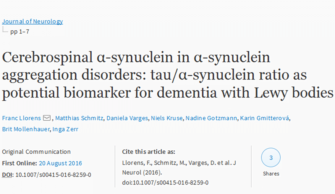 “Cerebrospinal α-synuclein in α-synuclein aggregation disorders: tau/α-synuclein ratio as potential biomarker for dementia with Lewy bodies” has been published in the Journal of Neurology. This research was supported in part by JPND through the DEMTEST project, selected in the 2011 biomarkers call.
