Author Archives: jpnd

Introducing the first global database of cohorts for neurodegenerative diseases

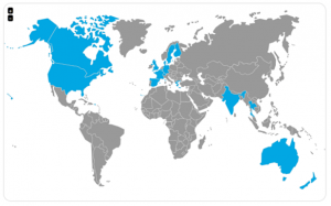
To promote the use and connection of cohort studies, the Joint Programme for Neurodegenerative Disease Research (JPND) has developed a new online gateway to longitudinal cohorts suitable for neurodegenerative disease (ND) research.

The JPND Global Cohort Portal is a searchable database of cohort studies from countries around the world. By providing users with an overview of and contact details for each cohort, this publicly available resource aims to facilitate information exchange and new collaborations, to help promote a greater volume of ND research, increased efficiency and ultimately greater scientific impact. The Portal is expected to debut on the JPND website in Autumn 2017 and will continue to grow as new cohorts are added.

How does it work?
The Portal spans both disease-focused and general population studies, providing a high-level overview of each cohort, including the types of data collected. Once the most relevant cohorts are identified, Portal users may navigate directly to the individual cohort website or contact the principal investigator for further information and guidance on how to use the cohort as a resource.

Why cohorts?
Longitudinal cohort studies allow researchers to collect data on populations over time in order to establish high-quality correlations and tease out new clues regarding the determinants and risk factors of disease. As such, cohorts are among the most important tools for current and future research on neurodegeneration.

As outlined in a 2013 JPND report linking and comparing studies could lead to important insights and statistically more powerful conclusions. Yet detailed information on cohort studies is often difficult to find, and identifying and connecting with the right contacts from each cohort can pose additional challenges. The JPND Global Cohort Portal was developed to meet these needs.

How can I get involved?
The JPND Global Cohort Portal will continue to expand. If you have information regarding a relevant cohort study that could be included, we encourage you to get in touch. For this and any other questions, emails should be directed to: [email protected].

Researchers have identified two groups of neurons that can be turned on and off to alleviate the movement-related symptoms of Parkinson’s disease. The activation of these cells in the basal ganglia relieves symptoms much longer than current therapies, like deep brain stimulation and pharmaceuticals.

The study was published in the journal Nature Neuroscience.

The study used optogenetics in a mouse model of Parkinson’s to better understand the neural circuitry involved in Parkinson’s disease, and could provide the basis for new experimental treatment protocols.

Parkinson’s disease is caused when the dopamine neurons that feed into the brain’s basal ganglia die and cause the basal ganglia to stop working, preventing the body from initiating voluntary movement.

The basal ganglia is the main clinical target for treating Parkinson’s disease, but currently used therapies do not offer long-term solutions.

To better understand how the neurons in the basal ganglia behave in Parkinson’s, the researchers looked at the inner circuitry of the basal ganglia. They chose to study one of the structures that makes up that region of the brain, a nucleus called the external globus pallidus (GPe). The GPe is known to contribute to suppressing motor pathways in the basal ganglia, but little is known about the individual types of neurons present in the GPe, their role in Parkinson’s disease or their therapeutic potential.

The research group used optogenetics, a technique that turns genetically tagged cells on and off with light. They targeted two cell types in a mouse model for Parkinson’s disease: PV-GPe neurons and Lhx6-GPe neurons. They found that by elevating the activity of PV-GPe neurons over the activity of the Lhx6-GPe neurons, they were able to stop aberrant neuronal behavior in the basal ganglia and restore movement in the mouse model for at least four hours — significantly longer than current treatments.

While optogenetics is used only in animal models, the researchers say their findings could lead to a new, more effective deep brain stimulation protocol.

Paper: “Cell-specific pallidal intervention induces long-lasting motor recovery in dopamine-depleted mice”
Reprinted from materials provided by Carnegie Mellon University.

A new large-scale genetic study found that low body mass index (BMI) is likely not a causal risk factor for Alzheimer’s disease, as earlier research had suggested, according to a study published in the Journal of Clinical Endocrinology & Metabolism.

To examine the association between Alzheimer’s disease and low BMI, the researchers analyzed blood and DNA samples from 95,578 participants in the Copenhagen General Population Study (CGPS). Of the participants, 645 individuals developed Alzheimer’s disease.

The researchers analyzed the study participants’ DNA for the presence of five genetic variants that have strong associations with BMI. Based on how many variants were found, participants were divided into four groups to reflect the likelihood of low BMI. The researchers also analyzed data from up to 249,796 individuals participating in the Genetic Investigation of ANthropometric Traits (GIANT) consortium for the genetic variants closely linked to low BMI.

The analysis found the presence of the genetic variants tied to low BMI was not associated with increased risk of Alzheimer’s disease. For comparison, the researchers examined if individuals with genetic variants connected to high BMI were more likely to have type 2 diabetes and did find the expected causal relationship.

Paper: “Body Mass Index and Risk of Alzheimer Disease: a Mendelian Randomization Study of 399,536 Individuals”
Reprinted from materials provided by The Endocrine Society.

A new study published in the journal Scientific Reports shows that bone marrow stem cell transplants helped improve motor functions and nervous system conditions in mice with amyotrophic lateral sclerosis (ALS) by repairing damage to the blood-spinal cord barrier.

The researchers say the results of their experiment are an early step in pursuing stem cells for potential repair of the blood-spinal cord barrier, which has been identified as key in the development of ALS.

Using stem cells harvested from human bone marrow, researchers transplanted cells into mice modeling ALS and already showing disease symptoms. The transplanted stem cells differentiated and attached to vascular walls of many capillaries, beginning the process of blood-spinal cord barrier repair.

The stem cell treatment delayed the progression of the disease and led to improved motor function in the mice, as well as increased motor neuron cell survival, the study reported. Because stem cells have the ability to develop into many different cell types in the body, researchers have focused on using stem cells to restore function lost through neurodegenerative disorders or injuries.

Damage to the barrier between the blood circulatory system and the central nervous system has been recently recognized as a factor in ALS development, leading researchers to work on targeting the barrier for repair as a potential strategy for ALS therapy.

In this study, the ALS mice were given intravenous treatments of one of three different doses of the bone marrow stem cells. Four weeks after treatment, the scientists determined improved motor function and enhanced motor neuron survival. The mice receiving the higher doses of stem cells fared better in the study.

The transplanted stem cells had differentiated into endothelial cells – which form the inner lining of a blood vessel, providing a barrier between blood and spinal cord tissue — and attached to capillaries in the spinal cord. Furthermore, the researchers observed reductions in activated glial cells, which contribute to inflammatory processes in ALS.

Paper: “Endothelial and Astrocytic Support by Human Bone Marrow Stem Cell Grafts into Symptomatic ALS Mice towards Blood-Spinal Cord Barrier Repair”
Reprinted from materials provided by USF Health.

Scientists have discovered a potential brain imaging predictor for dementia, which illustrates that changes to the brain’s structure may occur years prior to a diagnosis, even before individuals notice their own memory problems.

The study, published in the Neurobiology of Aging, looked at older adults who are living without assistance and who were unaware of any major memory problems, but scored below the normal benchmark on a dementia screening test.

Within these older adults, researchers also found evidence of less brain tissue in the same subregion of the brain where Alzheimer’s disease originates (the anterolateral entorhinal cortex located in the brain’s temporal lobe).

This is the first to measure this particular brain subregion in older adults who do not have a dementia diagnosis or memory problems that affect their day-to-day routine. It is also the first study to demonstrate that performance on the Montreal Cognitive Assessment (MoCA) dementia screening test is linked to the volume (size) of this subregion, along with other brain regions affected early in the course of Alzheimer’s disease.

The team studied 40 adults between the ages of 59 and 81 who live independently (or with a spouse) at home. All participants were tested on the MoCA. Those scoring below 26 — a score that indicates a potential problem in memory and thinking skills and suggests further dementia screening is needed — were compared to those scoring 26 and above.

Scientists were able to reliably measure the volume of the anterolateral entorhinal cortex by using high-resolution brain scans that were collected for each participant.

The strongest volume differences were found in the exact regions of the brain in which Alzheimer’s disease originates. The researchers are planning a follow-up study to determine whether the individuals who demonstrated poor thinking and memory abilities and smaller brain volumes indeed go on to develop dementia.

Paper: “Anterolateral entorhinal cortex volume predicted by altered intra-item configural processing”
Reprinted from materials provided by Baycrest Centre for Geriatric Care.

In experiments with a protein called Ephexin5 that appears to be elevated in the brain cells of Alzheimer’s disease patients and mouse models of the disease, researchers show that removing it prevents animals from developing Alzheimer’s characteristic memory losses. In a report on the studies, published in The Journal of Clinical Investigation, the researchers say the findings could eventually advance the development of drugs that target Ephexin5 to prevent or treat symptoms of the disorder.

Curious about whether Ephexin5 — a protein regulated by the protein EphB2 and thought to be responsible for inhibiting the development of dendritic spines — might play an important role in Alzheimer’s disease symptoms, researchers first investigated whether this protein might be poorly regulated in Alzheimer’s animal models and patients.

The researchers discovered that when they added amyloid beta to healthy mouse brain cells growing in petri dishes, these cells began overproducing Ephexin5. Additionally, when they injected the brains of healthy mice with amyloid beta, cells there also began overproducing Ephexin5 — both clues that the protein that makes Alzheimer’s characteristic plaques appears to trigger an increase in brain cells’ production of Ephexin5 of between 1- and 2.5-fold.

When the researchers examined preserved brain tissues isolated from Alzheimer’s patients during autopsies, they also found similarly high levels of Ephexin5. Additionally, they found elevated levels of Ephexin5 in mice genetically engineered to overproduce amyloid beta, and that show memory deficits similar to those with human Alzheimer’s disease, further confirming that excess Ephexin5 is associated with this disease.

Armed with what they called this wealth of evidence that brain cells produce too much Ephexin5 when Alzheimer’s disease linked to amyloid beta is present, the researchers then investigated whether reducing Ephexin5 might prevent Alzheimer’s deficits.

Using genetic engineering techniques that knocked out the gene that makes Ephexin5, the researchers developed mouse Alzheimer’s disease models whose brain cells could not produce the protein. Although the animals still developed the characteristic Alzheimer’s amyloid plaques, they didn’t lose excitatory synapses, retaining the same number as healthy animals as they aged.

To see whether this retention of excitatory synapses in turn affected behavior related to memory tasks, the researchers trained healthy mice, mouse models of Alzheimer’s and Alzheimer’s models genetically engineered to lack Ephexin5 in two learning tasks: one that involved the ability to distinguish objects that had moved upon subsequent visits to the same chamber, and another that involved the ability to avoid chambers where they’d previously received a small electric shock.

While the typical Alzheimer’s disease model mice appeared unable to remember the moved objects or the shocks, the Alzheimer’s animals genetically engineered to be Ephexin5-free performed as well as healthy animals on the two tasks.

The researchers caution that while the results all suggest removing Ephexin5 prevented Alzheimer’s disease-associated impairments, they don’t on their own provide a true test for the approach to treatment. That’s because in people with Alzheimer’s disease, the brain is exposed to amyloid beta for some time, probably decades, before any treatments might be administered.

To better reflect that human scenario, the researchers raised mouse models for Alzheimer’s disease into adulthood — allowing their brains to be exposed to excess amyloid beta for weeks — before injecting their brains with a short piece of genetic material that shut down Ephexin5 production. These mice performed just as well on the memory tasks as the healthy mice and those genetically engineered to produce no Ephexin5.

Together, these results suggest that too much Ephexin5 triggered by amyloid beta and reduced EphB2 signaling might be the reason why Alzheimer’s disease patients gradually lose their excitatory synapses, leading to memory loss — and that shutting down Ephexin5 production could slow or halt the disease.

Paper: “Reducing expression of synapse-restricting protein Ephexin5 ameliorates Alzheimer’s-like impairment in mice”
Reprinted from materials provided by Johns Hopkins Medicine.